Blogs
Ventilation Essentials: A Thorough Guide to Anatomy and Physiology
Date: November 29, 2023 | Posted by: Vikram Aditya Tirthani
Date: November 29, 2023 | Posted by: Vikram Aditya Tirthani
Embarking on a journey through the intricate landscape of ventilation requires unraveling the tapestry of anatomy and physiology. In this comprehensive guide, we will delve into the fundamentals of ventilation, exploring its definition, the mechanics of breathing, the key muscles orchestrating inspiration, and the intricate processes involved in gas exchange.
Ventilation is a dynamic physiological process that ensures the exchange of oxygen and carbon dioxide in the lungs via mechanical movement of gas or air into (inhalation) and out of (exhalation) the lungs
In other words it is a continual renewal of the air in the gas exchange areas of the lungs caused by the expansion & contraction of the chest cavity
Understanding the mechanics of breathing is fundamental to grasping the role of ventilators. The respiratory cycle involves two key phases: inhalation (inspiration) and exhalation (expiration). This rhythmic process ensures the exchange of gases necessary for sustaining life.
It involves below important factors –
· Major and accessory muscles
· Alveolar surface tension
· Elastic properties of the lungs and chest wall
· Resistance to airflow through the conducting airways

The duo leading the charge during inspiration includes the diaphragm and external intercostal muscles. As the diaphragm contracts, it flattens downward, creating a vacuum that draws air into the lungs. Simultaneously, the contraction of external intercostal muscles elevates the anterior portion of the ribs, expanding the thoracic cavity.
1. When the diaphragm contracts it flattens downwards
· Increasing the volume of the thoracic cavity
· Creating a negative pressure which assists to draw gas into the lungs
2. Contraction of External Intercostal muscles elevates the anterior portion of the ribs
· Increasing volume a little more
· Inspiration at rest is usually assisted by the diaphragm alone
(and essentially only muscle during REM Sleep)
While the diaphragm and external intercostals are primary players, certain situations call upon accessory muscles. The sternocleidomastoid, scalene, and pectoralis minor muscles lend their support during increased respiratory effort, ensuring a robust inhalation process.
· Sternocleidomastoid
· Scalene muscles
These muscles help to increase the thorax during inspiration when the minute volume is very high
· During exercise
· During increased work of breathing because of disease
Unlike inspiration, expiration is generally a passive process. Elastic recoil of the lungs and chest wall, coupled with the relaxation of the diaphragm and external intercostal muscles, facilitates the expulsion of air. There are no major muscles dedicated to expiration under normal circumstances.
· Expiration is passive and therefore requires no muscular effort
· Diaphragm relaxes
· The accessory muscles
· Abdominal
· Internal Intercostal muscles
· Assist expiration when the minute volume is high when expiration exceeds Functional Residual Capacity (FRC) or when airway obstruction is present
· Assist expiration for coughing and sneezing
· Inhalation brings in oxygen from the atmosphere into the lungs

The atmosphere contains:
· Approximately about 21% oxygen (O2)
· Almost no Carbon Dioxide (0.03% CO2), as this is a waste product from Respiration
· Exhalation expels Carbon Dioxide and other trace waste gases from the lungs
Regulation of ventilation –
· The aim is to maintain appropriate levels of oxygen & carbon dioxide in the blood
· Performed by the respiratory center
· Chemoreceptors detect changes in the blood gases, sending a message to the respiratory center, which then adjusts ventilation

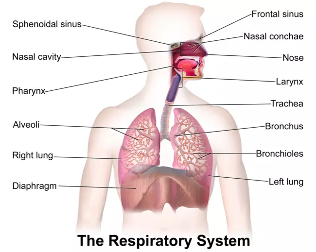
Ventilation traverses through the concept of dead space, encompassing parts of the respiratory system where air doesn’t participate in gas exchange. The trachea, bronchi, and bronchioles collectively form the conducting airways, highlighting the importance of optimizing oxygen delivery to the alveoli.
· With each breath, a portion of the tidal volume remains in the conducting airways.
· Does not reach the alveoli
· Does not participate in gas exchange
· Known as “dead-space ventilation”
· This normal dead space is also called Anatomical Dead Space
· In Disease states a portion of the alveoli and respiratory bronchioles do not participate in gas exchange
· Thus increasing the amount of dead-space ventilation
· The combination of this additional dead space and the anatomic dead space is called the physiologic dead space
The respiratory system unfolds into conducting and gas exchange airways. Conducting airways, comprising the trachea and bronchi, act as conduits for air. Gas exchange airways, encompassing the respiratory bronchioles and alveoli, facilitate the vital exchange of oxygen and carbon dioxide.

Clearance and filtering of foreign matter
· Coughing, gagging, sneezing
· Mucociliary transport system


Optimizes gas exchange and protects the delicate lung tissue
Mucociliary transport system consists of
· Ciliated Epithelial Cells
· Cells with cilia
· Aqueous Layer
· A thin layer of low-viscosity fluid (Sol)
· Gel Layer
· A mucous layer that floats on top of the aqueous layer
Terminal bronchioles divide to form the respiratory bronchioles
Gas exchange airways are made up
· Respiratory bronchioles
· Alveolar ducts
· Alveoli


Alveoli are spherical structures
· Have shared walls or septa
· Pores of Kohn permit some air to pass through the septa from the alveolus to the alveolus
· Promoting collateral ventilation and even distribution of air among the alveoli
· Consists of an epithelial layer and a thin elastic basement membrane
· Surfactant is a lipoprotein that coats the inner surface of the alveolus and facilitates expansion during inspiration
Gases are exchanged between the air and the blood (at tissue level) by diffusion
Gases exchanged are O2 and CO2
· O2 consumption about 3 ml /kg /min
· CO2 production 70% to 100% of O2 consumption
These are expressed as Partial Pressures or P
· Oxygen = PO2,
· Carbon Dioxide = PCO2


Oxygen:
· is transferred from the alveoli into the blood, & then by red blood cells to the various body tissues
· is used in the process of metabolism
Carbon Dioxide:
· is produced in the process of metabolism, enters the capillaries & is transported back to the site of gas exchange in the lungs
· is transported via red blood cells into the alveoli where it is expelled during exhalation.
The gas exchange process is achieved by –
1. Ventilation – The mechanical movement of air into and out of the lungs.
2. Diffusion – The movement of gases between air and spaces in the lungs and the bloodstream.
3. Perfusion – The movement of blood into and out of the capillary beds of the lungs to body organs and tissues – Cardiovascular system.
At the core of ventilation lies the gas exchange process, predominantly through diffusion. In the alveoli, oxygen seamlessly transitions from air sacs into the bloodstream, while carbon dioxide makes its journey in the opposite direction. This delicate dance is meticulously facilitated by ventilators to maintain optimal oxygen levels.
· Area of blood-gas barrier 50-100 m2
· The thickness of barrier < 0.5 micron
· CO2 diffuses about 20 faster than O2
· Red blood cell spends about 0.75 sec in pulmonary capillary normally 0.25 sec during exercise

Perfusion, the circulation of blood through the lungs, is integral to the gas exchange process. Ventilation ensures a harmonious interplay between oxygenated blood and tissues, while efficiently removing carbon dioxide from the bloodstream.
· Occurs across the Alveolocapillary membrane
· Normal perfusion approx. 100 ml of blood in the pulmonary capillary bed is spread over 70-100m2
· Any disorder that thickens the membrane impairs gas exchange
Ventilation, an intricate interplay of anatomy and physiology, emerges as a symphony of breath that sustains life. As we navigate through the complexities of the respiratory system, we gain a profound appreciation for the orchestrated precision required to harmonize the dance of oxygen and carbon dioxide within the human body. Ventilation stands as a testament to the marvels of medical science, where understanding the mechanics of breath opens the door to sustaining and enhancing life itself.
Get in touch with us using our contact form.